Entamoeba histolytica | Parasitology
Entamoeba histolytica | Parasitology
Entamoeba histolytica
Entamoeba histolytica is worldwide in distribution but more common in tropical and Subtropical countries. Trophozoites of Eh lives in the mucosa and submucosa of the large intestine of man.
 |
| Entamoeba histolytica | Medical Microbiology |
Morphology
The parasite exists in 3 forms:
- Trophozoite,
- precystic stage
- cyst
 |
| forms of Entamoeba histolytica |
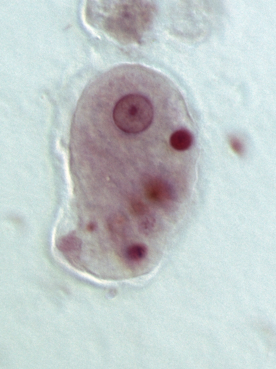
Trophozoite
- It measures 18-40 um in diameter. The Cytoplasm of the trophozoite is divided into 2portions
- (1) A clear outer ectoplasm (2) A granular endoplasm
- Trophozoites are motile by pseudopodia.
- Pseudopodia are long finger-like projections of Lectoplasm into which endoplasm flows.
- Trophozoites have a Nucleus of trophozoites that is spherical in shape and varies in size from 4-6 μm.
Pre-cystic Stage :
It is smaller in size and varies from 10-20 um in diameter. It is round or oval with blunt pseudopodia Its nucleus has a Similar character as that protozoan to cyst.
Cyst
It is round 10-15 Um in diameter and it is Surrounded by the cyst wall.
Life cycle :
- Entamoeba histolytica passes its life cycle only in one host, The man.
- The uninucleate trophozoite of Entamoeba histolytica occasionally inhibits the lower portions of the small intestine but it is Common in the Colon and rectum of humans.
- Within the host gut, the trophozoites asexually multiply fission.
- Trophozoites are active feeding from.
- Some of them invade the lining of the large intestine
- within the gut wall, some trophozoites are carried are away by the blood circulation to the liver lungs brain and other organs in which these amoebae are lodged and form the abscesses.
- From time to time, certain intestinal trophozoites assume the pre-cystic form
- At this stage. amoebae are preparing 'encyst'.
- This is the process of transformation of a trophozoite to a cyst
- This process of encystation takes place within few hours and the life span of the mature cyst inside. the host is only two days.
- These cysts are infective forms and are passed out of the body of host in faeces.
- The cysts are highly resistant to desiccation and Certain chemicals.
- The mature cyst has 4 (quart) nucleus
- Man acquire (get) the infection by ingestion. of water and food containing these cysts.
- When the cyst reaches the Caecum. or the lower part of the ileum
- The excystation occurs during this process.
- each mature Cyst librates a Single Amochae with 4 nuclear...
- which eventually produces 8 metacystic. trophozoites by the division of nuclei by binary fission.
- During growth Eh. Secrets a proteolytic enzyme that brings above destruction and necrosis of tissue leading to a flask-shaped ulcer.
- A large no of trophozoites are excreted along with blood and mucus in the faeces
- This condition is called an amoebic condition
 |
| The life cycle of Entamoeba histolytica |
Pathogenicity
- Entamoeba histolytica as the name suggests - histo-lytic = tissue destroying causes. internal amoebic dysentery and extraintestinal amoebiasis.
- During growth Eh. secrets a proteolytic ferment of the nature of histolysis, that brings about destruction. and necrosis of tissues. destruction
- This helps the parasite in obtaining nourishment
Laboratory Diagnosis :
Intestinal Amoebiaus
The incubation period is 1-4 weeks The Amoebae ulcerative lesions and profuse bloody diarrhoea. this is called acid Amoeboe dysentery.
Examination of Stool
- In acute amoebic dysentery, the stool is examined by necked eyes and microscopic Examination normal saline preparation is useful for the demonstration of actively motile trophozoites.
- While iodine preparation is required for the study of cyst dead trophozoites excretion of cyst in the Stool. is often intermitted.
- Therefore at least 3 days Consecutive specimen Should be examined.
- Charcol Layden Crystals may appear in saline preparation
- These are diamond Shaped crystals, clear and refractile.
- Amoebic dysentery must be differentiated from bacillary dysentery.
 |
| lab diagnosis of Entamoeba histolytica |
Extraintestinal Amoebians (Liver Abscess]
- Liver abscess is the most common manifestation of extraintestinal amoebiasis.
- An amoebic live abscess is associated with fever and abdominal pain in most patients.
- It occurs more commonly in adults than in children.
- Ultrasonography, MRI, and CT scan may be useful for the diagnosis.
Stool examination:
Cyst of Eh. are present in less than 15 per cent of Cases of amoebic liver abscesses and gives information regarding the persistence of intestinal infection.